Atherosclerosis is a leading cause of death worldwide. Currently, angiography is the primary clinical method for diagnosing atherosclerosis, but angiography can only assess lumenal occlusions. Increasing evidence indicates that it is atherosclerotic plaque rupture rather than severity of occlusion that leads to acute infarction or sudden cardiac death. However, sensitive detection and differentiation of vulnerableversus stable atherosclerotic plaques in vessels remains limited. There is evidence that composition and presence of specific markers can indicate if a plaque is at risk for rupture. Our research focuses on development of multifunctional nanoparticles targeted to atherosclerotic markers for multimodality imaging of atherosclerotic plaques.
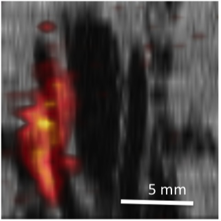
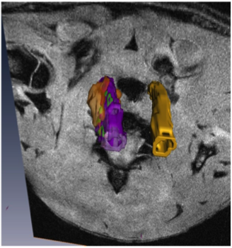
We have developed multimodal probes for PET/MR/optical imaging based on iron oxide nanoparticles or silicon quantum dots. A higher density of macrophages in plaques has been correlated with instability and localization to the plaque shoulders indicates greater risk of rupture. To image macrophage density we coat our particles with dextran sulfate, a ligand of macrophage scavenger receptor class A (SR-A). The highly sensitive positron emission tomography (PET) is used to identify putative plaques, high resolution magnetic resonance imaging (MRI) is then performed on these regions to obtain 3-dimensional images of the plaques and assess their vulnerability to rupture, and fluorescence allows for subsequent histology. In addition, as part of a Bioengineering Research Partnership awarded by NIH/NIBIB, we are also developing combined PET/MRI probes to support an instrument for simultaneous PET and MR imaging being developed by collaborators at UC Davis and Caltech